Classification: Homo sapiens Nine systems · 78 organs · 37 trillion cells
The Human Body
An annotated reference to the machine you live inside. Every plate is labelled, every figure sourced, and every explanation available in two registers — plain language, or clinical terminology.
System 01 · Skeletal systema skeletale
A frame that carries you — and rebuilds itself while you sleep.
Bone looks finished, but it isn’t. Cells tear it down and lay it back again continuously, so the skeleton you carry today is not the one you had a decade ago. It also stores minerals, manufactures blood, and absorbs the load of every step you take.
Osseous tissue undergoes continuous remodelling: osteoclasts resorb mineralised matrix while osteoblasts deposit osteoid, replacing roughly 10% of adult skeletal mass annually. The skeleton is the principal calcium–phosphate reservoir and houses haematopoietic marrow within trabecular compartments.
- 206
- Bones, adult
from ~270 at birth - 10%
- Remodelled
per year - 15%
- Of total
body mass
Source: Standring (ed.), Gray’s Anatomy, 42e, 2020
Sense of scale — where the bones are
Twenty-seven bones in each hand, twenty-six in each foot. Just over half the skeleton is given to grasping and walking.
System 02 · Muscular systema musculare
Six hundred engines, and every one of them can only pull.
Muscle never pushes. Each one shortens by up to a third of its length and then has to be pulled back by an opposing partner, which is why the body is built in pairs. Precision comes from how few fibres you recruit, not how many.
Skeletal muscle generates force through actin–myosin cross-bridge cycling; sarcomeres shorten up to ~30% of resting length. Movement arises from agonist–antagonist pairs acting across joints, with force graded by orderly motor-unit recruitment from small to large.
- 600
- Skeletal
muscles (approx.) - 40%
- Of body mass
upper range - 30%
- Max fibre
shortening
Source: Gray’s Anatomy, 42e · Guyton & Hall, 14e, 2021
Sense of scale — muscle against frame
The engine outweighs the chassis it moves by more than two to one.
System 03 · Nervous systema nervosum Deep dive
Eighty-six billion cells, arguing at 120 metres per second.
Your brain runs on about twenty watts — less than a household bulb — while handling balance, language, memory and grief at once. Signals race down insulated fibres as electrical pulses, then leap chemical gaps thinner than a wavelength of light.
Action potentials propagate saltatorily along myelinated Aα fibres at up to 120 m/s, then transduce chemically across synaptic clefts of 20–40 nm. The CNS accounts for ~2% of body mass yet ~20% of resting oxygen and glucose consumption.
- 86B
- Neurons
Azevedo, 2009 - 120
- m/s peak
conduction - 20W
- Resting power
draw, brain
Source: Azevedo et al., J. Comp. Neurol., 2009 · Guyton & Hall, 14e
Sense of scale — signal speed
A reflex reaches your spinal cord faster than any train you can buy a ticket for.
System 04 · Cardiovascular systema cardiovasculare
A pump the size of your fist, on a hundred-thousand-kilometre circuit.
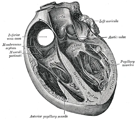
The heart is two pumps in one housing: the right side sends blood a short way to the lungs, the left drives it everywhere else. It repeats the cycle roughly once a second, without a single day off, for a lifetime.
The heart functions as a dual pump in series: the right ventricle perfuses the pulmonary circuit at ~25/10 mmHg, the left the systemic circuit at ~120/80 mmHg. Resting cardiac output is ~5 L/min (stroke volume ~70 mL × heart rate ~70 bpm).
- 5L
- Per minute
at rest - 100k
- km of vessels
estimated - 2.5B
- Beats in an
average life
Source: Guyton & Hall, 14e, 2021 · Gray’s Anatomy, 42e
Sense of scale — total vessel length
Laid end to end, the circuit would wrap the planet two and a half times.
System 05 · Respiratory systema respiratorium
Seventy square metres of surface, folded into your chest.
Lungs don’t breathe — they’re passive bags. The diaphragm drops, pressure inside the chest falls below the air outside, and the atmosphere is pulled in. Oxygen then crosses a barrier thinner than a soap film.
Ventilation is driven by diaphragmatic contraction generating sub-atmospheric intrapleural pressure. Gas exchange proceeds by passive diffusion across the alveolar–capillary membrane (~0.2–0.6 µm) along partial-pressure gradients.
- 480M
- Alveoli
Ochs, 2004 - 70m²
- Gas exchange
surface - 11k
- Litres of air
per day
Source: Ochs et al., Am. J. Respir. Crit. Care Med., 2004
Sense of scale — the tennis-court myth
Lungs are often said to equal a tennis court. They are closer to a third of one — which is still seventy square metres folded inside your ribs.
System 06 · Digestive systema digestorium
Nine metres of chemistry, turning matter into you.
The tract is one continuous tube from mouth to end — technically the outside world, threaded through you. Food only becomes part of your body once it has been broken small enough to cross that wall, a journey of one to three days.
The alimentary canal is a continuous mucosa-lined tube; absorption occurs chiefly in the jejunum, where plicae, villi and microvilli amplify surface area 60–120×. Roughly 5×10⁸ enteric neurons coordinate motility semi-autonomously of the CNS.
- 9m
- Tract length
mouth to anus - 30m²
- Absorptive area
Helander, 2014 - 24–72
- Hours total
transit time
Source: Helander & Fändriks, Scand. J. Gastroenterol., 2014 · Sender et al., 2016
Sense of scale — the tennis-court myth, again
Helander and Fändriks measured the real figure at about thirty square metres — nearer half a badminton court than a tennis court.
System 07 · Sensory systema sensorium
Your retina is not part of your eye. It is brain that grew out to meet the light.
The retina isn’t a sensor wired into the brain — it is brain. Early on, a piece of the developing forebrain pushes outward, cups over on itself, and becomes the light-sensitive lining at the back of the eye. It is the only stretch of central nervous system anyone can see without cutting you open, which is what an optician is actually looking at.
The neural retina arises as an evagination of the diencephalon: the optic vesicle invaginates to form the optic cup, whose inner layer becomes the neural retina and outer layer the retinal pigment epithelium. The optic nerve is therefore a CNS tract myelinated by oligodendrocytes, not a peripheral nerve.
- 120M
- Rods
mostly peripheral - 6.5M
- Cones
massed at the fovea - 0
- Photoreceptors
at the optic disc
Source: Purves et al., Neuroscience, NCBI · StatPearls, Embryology — Optic Cup
System 08 · Urinary systema urinarium
They throw away a hundred and eighty litres a day, then take back ninety-nine per cent of it.
Kidneys don’t strain waste out of blood. They dump nearly everything through a filter — water, salt, sugar, amino acids — and then reclaim almost all of it, keeping back only what is genuinely surplus. A hundred and eighty litres go through each day; one or two leave the body. It looks absurdly wasteful, and it is exactly what makes the control so fine.
Glomerular filtration proceeds at roughly 120 mL/min, some 180 L/day. Approximately 99% of the filtrate is reabsorbed across the proximal tubule, loop of Henle, distal tubule and collecting duct, yielding 1–2 L of urine daily. The kidneys receive 20–25% of resting cardiac output.
- 180L
- Filtered
every day - 99%
- Reabsorbed
before it leaves - 1M
- Nephrons
per kidney
Source: StatPearls, Physiology — Glomerular Filtration Rate · Histology, Nephron
System 09 · Integumentary systema integumentarium
The part of you that touches the world is already dead.
Every handshake, every gust of wind, every cold doorknob — you meet all of it through fifteen to twenty layers of dead cells. The outer skin is built from cells that climbed up from below, flattened, threw out their own nucleus and died on purpose. That dead sheet is the whole barrier between you and everything else, and it is rebuilt continuously from underneath.
The stratum corneum comprises 15–20 layers of anucleate corneocytes, the terminal differentiation product of epidermal keratinocytes. Cells migrate from the stratum basale to the corneum over approximately 28 days; complete epidermal turnover is estimated at 40–56 days in humans. The skin is the largest organ of the body.
- 15–20
- Layers of dead cells
you present outward - 28
- Days from the base
to the surface - 40–56
- Days for the whole
epidermis to turn over
Source: Making an Epidermis, NIH · Dissecting the formation, structure and barrier function of the stratum corneum
Section 10 · Connections Part two
Nine systems is a filing convenience. The body has never observed it.
Dividing the body into systems is how it gets taught, not how it works. The most interesting structures belong to two or three at once, and the divisions fall apart exactly where they are load-bearing. Six places the boundaries fail.
-
02Muscular 05Respiratory 03Nervous
The diaphragm answers to two masters
It is skeletal muscle — the voluntary kind — which is why you can hold your breath. But it is also the only muscle you cannot abandon: the brainstem drives it whether or not you attend to it, about 20,000 times a day. It fails upward through the neck, not the chest, because it forms early and migrates down, dragging its nerve behind it.
C3–C5Phrenic nerve roots. A neck injury above them stops breathing; below them does not.
Source: Gray’s Anatomy, 42e · Guyton & Hall, 14e
-
Your bones make your blood
The skeleton is not scaffolding with marrow stored inside it — the marrow is the point. Red marrow in the pelvis, sternum, ribs and vertebrae runs the body’s largest manufacturing operation, and it never stops, because every cell it makes is on a countdown from the day it is released.
2M/secRed cells produced, each lasting 100–120 days before the spleen retires it.
Source: Chen et al., Front. Endocrinol., 2018 · Gray’s Anatomy, 42e
-
01Skeletal 02Muscular 04Cardiovascular
The skeleton is a bank, and muscle spends the deposit
Calcium is what lets a muscle fibre contract at all, and what lets the heart keep its rhythm. Almost all of it is locked in bone as hydroxyapatite — which means your skeleton is not only holding you up, it is underwriting every heartbeat. When blood calcium falls, the body will dismantle bone to defend it, because the rhythm matters more than the frame.
99%Of body calcium held in bone, drawn on by PTH, calcitonin and calcitriol.
Source: StatPearls, Physiology — Calcium, 2023
-
Your gut has its own brain, and does not consult you
Embedded in the wall of the intestine is a nervous system large enough to run the tract by itself. Sever every connection to the brain and the gut carries on digesting — coordinating peristalsis, secretion and blood flow without instruction. It is the only organ that would keep working if you cut it off from the central nervous system entirely.
5×10⁸Enteric neurons, coordinating motility semi-autonomously of the CNS.
Source: Gray’s Anatomy, 42e · Guyton & Hall, 14e
-
05Respiratory 04Cardiovascular
Two systems meet across a film you could see through
The whole purpose of breathing and the whole purpose of circulation converge at one boundary: the sheet where air and blood are separated by almost nothing. No pump pushes oxygen across it. It moves by diffusion alone, down a pressure gradient, over a distance shorter than a wavelength of visible light.
0.2–0.6µmAlveolar–capillary membrane. Seventy square metres of it, folded into the chest.
Source: Ochs et al., Am. J. Respir. Crit. Care Med., 2004
-
The heart cannot get blood back from your feet
Arteries have the pump behind them. Veins do not — by the time blood reaches your ankles the pressure from the heart is largely spent, and it still has to climb back up. What lifts it is your calf muscles squeezing the veins between them, with one-way valves keeping each squeeze. Standing perfectly still is the one posture that defeats it, which is why soldiers on parade faint.
64%Of total blood volume sitting in the systemic veins at rest, waiting to be moved.
Source: Guyton & Hall, 14e, 2021
Part Three
When something goes wrong
Three ordinary complaints, traced back to the mechanism that produces them. These pages explain how a symptom is generated — they do not diagnose one. Nothing here is a substitute for a clinician.
Condition 11 · Headache cephalalgia
The brain cannot feel pain. A headache is everything around it complaining.
Cut into brain tissue and it registers nothing — there are no pain receptors in it at all. Surgeons operate on a conscious brain for exactly this reason. What hurts is the packaging: the tough membrane wrapping the brain, the big arteries at its base, and the muscles and vessels of the scalp outside the skull.
The cranium, brain parenchyma, ependymal lining and choroid plexus are pain-insensitive. Nociception arises from the dura mater and dural arteries, the proximal arteries of the circle of Willis, the great venous sinuses, cranial nerves, and extracranial muscle and vasculature — transmitted largely via trigeminal afferents with peripheral and central sensitisation.
- 40%
- Of people affected
3.1 billion, 2021 - 70%
- Report episodic
tension-type - 3rd
- Migraine rank,
global DALYs
Source: WHO headache disorders, 2024 · Clinical Methods, 3e, ch. 59
- 01The insensitive core. Skull, brain tissue, the ventricle lining and choroid plexus carry no nociceptors. Damage there produces no sensation of its own.
- 02The sensitive wrapper. The dura and its arteries, the large vessels at the base of the brain and the great venous sinuses are richly supplied with pain fibres.
- 03Trigeminal alarm. Dural nerve endings release CGRP and substance P, widening vessels and inflaming the membrane. The fibres then become easier to trigger than before.
- 04The outside layer. In tension-type headache, sustained contraction of scalp and neck muscles adds a second source — which is why the ache feels like a band rather than a point.
Condition 12 · Abdominal pain dolor abdominis
Your gut cannot feel a cut. It can only feel a stretch.
The nerves in your intestines ignore things that would be agony on skin. You can cut or burn the bowel wall and feel nothing. What they do report is stretching — a tube being pulled wider than it should be. That is why a stomachache is a vague, deep, hard-to-point-at ache rather than a sharp spot.
Visceral nociceptors respond to stretch as the primary stimulus, along with distension, contraction, traction, torsion and ischaemia, but not to cutting or thermal injury. Afferents travel in unmyelinated C-fibres entering the cord bilaterally at multiple levels, producing dull, poorly localised midline pain.
- C
- Unmyelinated fibres
slow, diffuse - 3
- Gut divisions
fore, mid, hind - Aδ
- Fibres that localise
once lining inflames
Source: StatPearls, Acute Abdomen, 2024 · Gray’s Anatomy, 42e
- 01A tube is stretched. Gas, obstruction or inflammation distends a hollow organ. Stretch — not cutting — is what the receptors in the wall are built to detect.
- 02The signal arrives everywhere at once. Slow C-fibres enter the spinal cord at several levels on both sides, so the brain gets no clear return address.
- 03The brain guesses the body wall. Gut and skin nerves converge on the same cord segments, so the ache is felt on the surface — foregut at the stomach, midgut at the navel, hindgut low down.
- 04Then it sharpens and moves. Once inflammation reaches the abdominal lining, fast Aδ fibres take over and pin it precisely — the reason appendicitis starts at the navel and ends in the right lower corner.
Condition 13 · Common cold rhinitis acuta
The symptoms are your defence, not the damage.
A cold virus barely harms you. Unlike flu, it does not wreck the lining of your nose — it slips into a few cells and copies itself. Almost everything you feel is your own immune system responding: the streaming, the blocked nose, the sore throat. You are not feeling the virus. You are feeling the reaction to it.
Rhinovirus binds ICAM-1 on nasal epithelium but, in contrast to influenza, produces no significant direct cytotoxicity. Symptoms derive from the innate immune response — bradykinin generation drives sore throat, rhinorrhoea and congestion, with interleukins and prostaglandins contributing systemic features.
- 2–5
- Colds per year
adult average - 160
- Rhinovirus subtypes
and counting - 1–4
- Days incubation
before symptoms
Source: StatPearls, Upper Respiratory Tract Infections, 2023 · NHS, Common cold
- 01The virus docks. Rhinovirus latches onto ICAM-1, a protein already sitting on your nasal cells, and gets inside. One to four days pass before you notice anything.
- 02Almost nothing is destroyed. Unlike influenza, rhinovirus causes no meaningful cell death. If damage caused symptoms, you would have none.
- 03The response arrives. Infected cells summon the innate immune system. Bradykinin produces the sore throat and the running nose; interleukins and prostaglandins produce the ache and fever.
- 04The block is blood, not snot. Veins in the turbinates dilate and engorge, narrowing the airway. That is why blowing your nose does not clear it — and why decongestants target vessels.
Condition 14 · Fever pyrexia
You shiver because the thermostat moved, not because you are cold.
A fever isn’t heat leaking in from an infection. Your body has a target temperature, and infection moves the target upward. The moment it does, your actual temperature — unchanged, still perfectly normal — reads as too low. So you shiver and reach for a blanket while running hot, which is the strangest part of being ill: you feel freezing precisely because you are burning.
Exogenous pyrogens induce endogenous pyrogens — IL-1, IL-6 and TNF-α — which act via the organum vasculosum of the lamina terminalis to raise prostaglandin E₂ in the preoptic area. PGE₂ binding EP3 receptors silences tonically inhibitory median preoptic neurons, disinhibiting the thermogenic circuitry and driving shivering and vasoconstriction until core temperature meets the elevated set point.
- PGE₂
- The final
mediator - EP3
- The receptor
it acts on - 38°C
- Usual clinical
threshold
Source: StatPearls, Physiology — Fever · Neural Mechanisms of Inflammation-Induced Fever, NIH
- 01A target exists. The preoptic hypothalamus holds a set point and defends it, the way a thermostat defends a room.
- 02Infection moves it. Pyrogenic cytokines raise prostaglandin E₂ in the preoptic area, and the set point is reset upward.
- 03Normal now reads as cold. Your temperature has not changed yet, but against the new target it is a deficit — so you feel freezing.
- 04The body closes the gap. Shivering and vasoconstriction generate and conserve heat until core temperature reaches the new set point. Then the chills stop.
Condition 15 · Bruise ecchymosis
A bruise changes colour because you are watching haemoglobin be taken apart.
The colours of a bruise are not the injury healing over. They are a chemical sequence, visible through your skin: the red of spilled blood, then the green and yellow of the pigment that carries oxygen being dismantled into its waste products and carried away. You are watching a demolition, in order, from the outside.
Extravasated erythrocytes lyse and haemoglobin is degraded by haem oxygenase to biliverdin, then rapidly to bilirubin, with residual iron stored as haemosiderin. Each intermediate has its own chromophore: haemoglobin red-blue, biliverdin green, bilirubin yellow, haemosiderin golden-brown. Products clear via lymphatic drainage, typically over about two weeks.
- 4
- Pigments,
in sequence - 5–10
- Days to the
green-yellow stage - 2wk
- Typical time
to clear
Source: Natural History of the Bruise, NIH · Hemoglobin-related cutaneous discoloration, NIH
- 01Blood leaves the vessel. A blow ruptures small vessels under intact skin. The trapped red cells are what you first see — red, darkening to blue-purple.
- 02Haem oxygenase starts work. The red cells lyse and haemoglobin is broken down to biliverdin, which is green. This is why a bruise turns a colour blood never was.
- 03Green becomes yellow. Biliverdin converts rapidly to bilirubin — the same pigment that yellows the skin in jaundice. Typically visible days five to ten.
- 04The iron is filed away. Residual iron is stored as golden-brown haemosiderin, and the products drain away through the lymphatics over roughly two weeks.
Section 16 · Live Running
While you have been reading.
Every rate below is one already sourced elsewhere on this page, running forward from the moment you arrived. Nothing here is measured from you. These are population averages doing arithmetic against a clock — an ordinary adult at rest, not a reading of your body.
- 0 Heartbeats At a resting 70 a minute
- 0 Breaths Some 20,000 a day
- 0 Red cells made Two million every second
- 0 Litres pumped Five a minute at rest
Rates: Guyton & Hall, 14e · Chen et al., Front. Endocrinol., 2018
Section 17 · Matter materia Coda
By weight you are mostly oxygen. By count you are mostly hydrogen.
Put a person on a scale and the largest single thing they are made of is oxygen — most of it locked inside water. Count the atoms instead and the answer flips entirely: hydrogen wins, and not narrowly, because a hydrogen atom weighs about a sixteenth of an oxygen one. Two true answers to the same question, and the only difference is whether you were counting or weighing.
Elemental composition differs markedly by basis of measurement. By mass, carbon, hydrogen, nitrogen and oxygen constitute 96.5% of body weight; adding phosphorus and calcium brings six elements to roughly 99%. By atom count hydrogen predominates, reflecting a relative atomic mass of 1 against oxygen’s 16 — water alone, at some 70% of cell weight, contributes two hydrogen atoms for every oxygen.
- 96.5%
- Of your weight
is four elements - 70%
- Of a cell’s weight
is water - 6
- Elements make up
about 99% of you
Source: Alberts et al., Molecular Biology of the Cell — The Chemical Components of a Cell, NCBI
Section 18 · Provenance unde scimus Coda
These plates required bodies. Almost none were given freely.
The engravings on this page are beautiful, and each one exists because somebody was dissected. From 1752 English law handed anatomists the bodies of executed murderers — dissection was written into the sentence, an extra punishment stacked on hanging, designed to deny the condemned a burial. It supplied about four bodies a year against schools that wanted hundreds. The shortfall was met by men who dug up the newly buried, and in Edinburgh by two who skipped the digging and murdered sixteen people. The Anatomy Act of 1832 ended the grave robbing by redirecting the supply to the workhouse dead — which moved it from the criminal poor to the merely poor.
The Murder Act 1752 designated post-mortem dissection as a component of capital sentencing, transferring the corpses of executed murderers to the Company of Surgeons. Legal supply remained an order of magnitude below demand as anatomical teaching expanded, sustaining an illicit trade in exhumed bodies and, in the Burke and Hare case of 1828, sixteen homicides committed for sale. The Anatomy Act 1832 legalised the use of bodies unclaimed 48 hours after death in workhouses, hospitals and prisons.
- 1752
- Dissection added
to the sentence - 4
- Bodies a year
it lawfully supplied - 1832
- Supply moved to
the unclaimed poor
Source: UK Parliament, Body snatching · Harnessing the Power of the Criminal Corpse, NCBI · Royal College of Surgeons
Section 19 · References
Every figure on this site traces back to a printed source.
Anatomical counts vary between authorities and across individuals. Where estimates differ, the figure quoted is the one most commonly reported in current reference texts, with the primary study named.
- 01Standring S. (ed.). Gray’s Anatomy: The Anatomical Basis of Clinical Practice, 42nd ed. Elsevier, 2020.
- 02Hall J.E. Guyton & Hall Textbook of Medical Physiology, 14th ed. Elsevier, 2021.
- 03Azevedo F.A.C. et al. Equal numbers of neuronal and non-neuronal cells make the human brain an isometrically scaled-up primate brain. J. Comp. Neurol., 2009.
- 04Ochs M. et al. The number of alveoli in the human lung. Am. J. Respir. Crit. Care Med., 2004.
- 05Helander H.F. & Fändriks L. Surface area of the digestive tract — revisited. Scand. J. Gastroenterol., 2014.
- 06Sender R., Fuchs S., Milo R. Revised estimates for the number of human and bacteria cells in the body. PLoS Biol., 2016.
- 07Blanda M. Headache. In: Walker H.K. et al. (eds). Clinical Methods: The History, Physical, and Laboratory Examinations, 3rd ed. Butterworths, 1990. NCBI Bookshelf.
- 08World Health Organization. Headache disorders — fact sheet. WHO, 2024. who.int.
- 09Patterson J.W. et al. Acute Abdomen. StatPearls. StatPearls Publishing, 2024. NCBI Bookshelf.
- 10Thomas M., Bomar P.A. Upper Respiratory Tract Infection. StatPearls. StatPearls Publishing, 2023. NCBI Bookshelf.
- 18Koster M.I. Making an Epidermis. Ann. N.Y. Acad. Sci., 2009; with Harding C.R. et al., Dissecting the formation, structure and barrier function of the stratum corneum. PMC2861991.
- 17Hurren E. et al. Harnessing the Power of the Criminal Corpse; and Dissecting the Criminal Corpse. Palgrave, via NCBI Bookshelf. NCBI Bookshelf. With UK Parliament, Body snatching, and the Royal College of Surgeons archives.
- 15Physiology, Fever. StatPearls. StatPearls Publishing. NCBI Bookshelf.
- 16Natural History of the Bruise: Formation, Elimination, and Biological Effects of Oxidized Haemoglobin. NIH / PMC. PMC3671564.
- 14Alberts B. et al. The Chemical Components of a Cell. In: Molecular Biology of the Cell, 4th ed. Garland Science, 2002. NCBI Bookshelf.
- 12Purves D. et al. (eds). Neuroscience, 2nd ed. Sinauer, 2001. NCBI Bookshelf.
- 13Embryology, Optic Cup; Physiology, Glomerular Filtration Rate; Histology, Nephron. StatPearls. StatPearls Publishing. NCBI Bookshelf.
- 11National Health Service. Headaches and Common cold — condition guidance. NHS, 2024. nhs.uk.